Three-dimensional mammography used in conjunction with conventional mammography reduced the rate of false alarms by more than a third compared to women who underwent traditional mammography alone, according to a new study from Yale School of Medicine published in the January issue of the journal Radiology.
The study provides further evidence that the advanced technology, also known as breast digital tomosynthesis, represents the “evolution of mammography” for breast cancer screening, said Dr. Melissa Durand, assistant professor of diagnostic radiology at Yale University School of Medicine and one of the study’s authors.
“With 3D mammography, women won’t have to experience the anxiety of a call back (for further testing) as often and at the same time they can feel more comfortable that their screening exam will find the types of cancers we worry about,” she said.
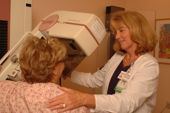
Photo Courtesy Of St. Francis Hospital and Medical Center.
Donna Wirthman, breast imaging supervisor at the Hoffman Breast Health Center, assists a patient undergoing 3D mammography.
Unlike conventional mammography that produces two-dimensional images, tomosynthesis produces a series of 3D images that display breast tissue in 1-millimeter sections. The technique reduces the superimposition of breast tissue, which can help radiologists differentiate between abnormalities and normal tissue, thereby decreasing the need for women to return for additional tests.
The Yale study looked at 17,955 screening mammograms. Forty-eight percent of the women had tomosynthesis and the remainder had two-dimensional mammography alone.
Among the findings:
• The rate of recalls – the number of women called back for additional testing because of a questionable finding – was 36.6 percent lower among women who had tomosynthesis when compared to women who had only conventional mammography.
• The recall rate was significantly lower for patients who had breast asymmetries and calcifications. The 3D images allow radiologists to determine that some breast asymmetries are overlapping tissue, rather than suspicious abnormalities, Durand explained.
• Cancer detection was slightly higher in the group of women who had tomosynthesis.
The Radiology study represents the latest scientific findings examining the effectiveness of 3D mammography, which an increasing number of health care providers across Connecticut and the nation are now offering patients.
Last year, JAMA published the largest study of its kind involving radiologists from academic and community-based practices nationwide who reviewed mammograms of nearly half a million women. The study found 3D mammography was associated with a decrease in the number of recalls and increased cancer detection. Yale took part in the JAMA study.
“Three-dimensional mammography is the only method for breast cancer screening that has demonstrated this dual benefit,” said Durand. Yale offers tomosynthesis to all patients regardless of age, breast density and risk factors.
At Stamford Hospital, the recall rate has decreased by 40 percent and cancer detection rates are 60 percent higher than two years ago, said Dr. David Gruen, director of Women’s Imaging. Stamford Hospital offers 3D mammography to all women regardless of breast density.
“We’re literally calling back thousands fewer women for unnecessary tests,” he said. “Our cancer detection rates are among the highest in the country. We think that’s important because Connecticut has the highest incidence of breast cancer in the country.”
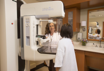
Photo Courtesy Of Stamford Hospital.
Mammography technologist Yolanda Jerez assists a patient undergoing 3D mammography at Stamford Hospital.
The “positive predictive value of biopsies” has also improved, meaning fewer women are undergoing unnecessary biopsies. “We’re taking biopsies of the right things,” he said.
Despite the evidence, some breast health experts continue to view 3D mammography as an investigational tool that warrants further study. They question its cost-effectiveness and worry about the higher dose of radiation associated with tomosynthesis.
The radiation dose for tomosynthesis is higher than for conventional mammography alone, but the dose is still below the safety limits set by the U.S. Food and Drug Administration, noted Durand. Computer software that would reconstruct the two-dimensional images from the 3D images would reduce the overall radiation dosage of tomosynthesis.
For Dr. Niamey Wilson, co-director of the Hoffman Breast Health Center at Saint Francis Hospital and Medical Center in Hartford, the “benefits outweigh the risks” when it comes to 3D mammography. She has seen a decrease in recalls and better cancer detection rates among women using tomosynthesis at Saint Francis, which offers the technology to patients with dense breasts, calcifications, and a high risk for breast cancer due to personal or family history.
“Tomosynthesis is one of the best improvements in screening mammography that we have seen in a long time,” said Wilson, a breast surgeon. “Our experience corresponds with the latest study out of Yale and reinforces what many people believe about the benefits of tomosynthesis.”
She pointed to a January 2015 study in the Journal of ClinicoEconomics and Outcomes Research that found the savings associated with follow-up services for each woman who underwent tomosynthesis was $28.53. That translates to a 20 cents savings per woman per month across the health plan’s membership and an overall cost savings to the plan of $2.4 million per year.
“This has immense potential cost savings for United States health care,” said Wilson, noting that an estimated 39 million mammograms are performed nationwide each year.
Durand expects the use of tomosynthesis to expand now that the Centers for Medicare and Medicaid Services began reimbursing health care providers for the procedure as of Jan. 1, 2015. The FDA also approved a second manufacturer of 3D mammography units. Until recently, facilities could only purchase the technology from a single vendor.
“I think more and more sites will acquire tomosynthesis in the near future in much the same way that digital mammography overtook film mammography in the mid 2000s,” she said. “This is the next step in breast imaging.”